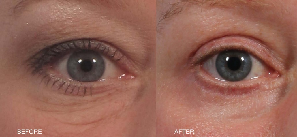
What it treats
Loose lower eyelid skin (lower dermatochalasis).
Why an eyelid skin pinch is used
Skin pinch blepharoplasty removes loose or redundant lower eyelid skin that cannot be sufficiently improved with non-surgical means (chemical peels, lasers, topicals). It removes skin only and typically does not touch the underlying muscle or orbital septum. This approach is thought to avoid some of the problems associated with surgery that does violate the muscle and orbital septum which has been associated with a higher rate of lower eyelid retraction or pulling down of the lower eyelid due to scar formation in these deeper eyelid layers.
How lower lid skin pinch is performed
Incision planning
The term skin pinch originates from the manner in which the procedure is planned. A tweezers or forceps are used by the surgeon to pinch the excess lower eyelid skin, just below the eyelashes. Sometimes this is done with the patient in a seated position to determine the desired contour of the eyelid, taking gravity into account.
As the surgeon pinches the extra skin, the position and shape of the lower eyelid should remain unchanged. If the eyelid is being pulled downward, less skin removal should be planned. If the lower eyelid is loose or poorly supported at its outer corner (ectropion or lower eyelid laxity) a canthopexy or canthoplasty should be performed at the same time to tighten and support the lower eyelid.
Typically, a marking pen is used to delineate the area for removal, extending horizontally, beneath the eyelash line. The upper edge of the marking should correspond with the lower eyelid crease if one is present. The outer edge of the markings may extend into a smile line.
Local anesthetic
A numbing fluid combined with a vasoconstrictor is injected with a thin needle beneath the lower eyelid skin for comfort and to reduce bleeding.
Incision and skin removal
The marked skin area is incised and removed, using either a scalpel, scissors, laser, electrosurgical instrument, or radiofrequency. The plane of dissection is just beneath the eyelid skin, on top of the orbicularis muscle. Cautery is performed as needed to ensure the site is bloodless.
Eyelid tightening (canthopexy) and orbicularis muscle tightening is then performed if required.
Closure
Small stitches are used to reapproximate the skin edges.
Follow-up care
An antibiotic ointment is applied periodically to the incision line until sutures are removed at 5 – 8 days.

