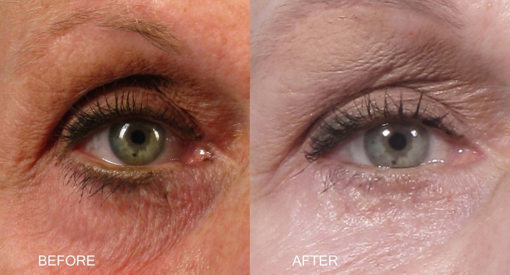
What it treats
These procedures provide support to the lower eyelid. They are also used to tighten a loose or sagging lower eyelid, improve the shape or position of the lower eyelid, or to re-attach a displaced lateral canthal tendon.
What is the difference between canthopexy and canthoplasty
The root “cantho” refers to the canthus, or the corner of the eyelids. There is an inner and outer canthus, or eyelid angle. Lateral canthopexy and canthoplasty (at the outer corner of the eyelids) is more common than medial procedures (at the inner corner). “Pexy” means that a structure is being fixated and “plasty” means that a structure is being modified.
With a lateral canthopexy, stitches are used to tighten or support the outer corner of the lower eyelid without severing the lateral canthal tendon or the outer eyelid angle. In a sense, the structures of the eyelid are not rearranged, they are just tightened “as is”. Theoretically with a canthopexy there is less risk of rounding the outer angle of the eyelids. Canthopexy is frequently performed with cosmetic lower eyelid surgery.
Lateral canthoplasty does un-attach the lateral canthal tendon and separate the angle at the outer corner of the eyelids. The tendon is then re-attached in order to tighten and/or reposition the lower eyelid orientation. Canthoplasty is generally more powerful than canthopexy, but may involve more risk of rounding at the outer eyelid angle.
How lower eyelid tightening procedures are done
Canthopexy
The area is numbed with an injection of anesthetic under the eyelid skin. An incision is made at the outer edge of the eyelid, beneath the eyelash line and below the outer corner of the eyelids extending into a skin crease. The length of this incision is typically 1 – 2 centimeters. Dissection is carried through the eyelid muscle (orbicularis oculus) to expose a section of the lower eyelid tarsus (a connective tissue structural plate of the eyelid) and the bony covering (periosteum) at the outer orbital rim. Stitches (usually permanent material) are placed to fasten and advance the tarsus to the inner aspect of the bony rim. The skin incision is closed with additional stitches.
Canthoplasty
An anesthetic injection with a small needle numbs the area at the outer corner of the eyelids. The lateral canthal angle, where the upper and lower eyelid meet at the outer corner, is horizontally separated. A lower eyelid attachment, the inferior crus of the lateral canthal tendon, is sharply released. A wedge of eyelid is removed in cases where the lower eyelid is being shortened. A tongue is created from the lateral edge of the lower eyelid tarsus by separating and removing the surrounding tissues. This tarsal tongue, or strip, is stitched to the inside edge of the orbital rim. Permanent or dissolvable stitches are used, and the strip is attached to the periosteum (the bony covering). A stitch is used to reform the lateral canthal angle, and stitches are used to close the skin incision after trimming any excess skin.
Follow-up care
Topical ophthalmic antibiotic ointment is applied with regular frequency after the procedure until the stitches are removed, typically at 5 to 10 days. Swelling and/or bruising is commonly seen during the days after the procedure. Complete healing can take several months.
Potential risks
These include rounding of the outer corner of the eyelids, recurrent sagging of the lower eyelid or incomplete improvement, visible scars, dry eyes, tearing, pulling down of the lower eyelid or eyelid retraction, and eyelid asymmetry.

